Abdominal hysterectomy
Abdominal Hysterectomy: Surgical Procedure and Overview
An abdominal hysterectomy is a surgical procedure involving the removal of the uterus through an incision made in the abdominal wall. This procedure is commonly performed to treat various medical conditions such as uterine fibroids, endometriosis, pelvic organ prolapse, and certain gynecological cancers. It can be performed for both benign and malignant conditions, depending on the patient’s medical history and diagnosis.
Procedure:
-
Preparation: Before the surgery, the patient is thoroughly evaluated through medical history, physical examination, and possibly imaging tests such as ultrasound or MRI. The surgical team will also conduct necessary preoperative tests to ensure the patient is fit for surgery.
-
Anesthesia: The patient is administered general anesthesia, which induces a deep sleep-like state and ensures they do not feel any pain during the procedure.
-
Incision: An incision is made in the abdominal wall, typically horizontally across the lower abdomen (bikini line incision) or vertically from the belly button to the pubic bone. The choice of incision type depends on the patient’s condition, surgeon’s preference, and the extent of the procedure.
-
Access and Exploration: After making the incision, the surgeon gains access to the abdominal cavity. The surrounding organs are carefully moved aside to expose the uterus and its supporting structures.
-
Uterine Manipulation: The blood vessels and ligaments that attach the uterus to the surrounding structures are identified, clamped, and ligated (tied off) to prevent excessive bleeding. The surgeon then carefully detaches the uterus from its attachments, including the cervix if necessary.
-
Removal: Once the uterus is detached, it is lifted out of the abdominal cavity through the incision. In some cases, the ovaries and fallopian tubes may also be removed (salpingo-oophorectomy) if they are diseased or if the patient’s condition requires it.
-
Closure: After the uterus is removed, the surgeon closes the incision using stitches or sutures. Depending on the incision type and surgeon’s preference, absorbable sutures or staples may be used.
-
Recovery: The patient is closely monitored in the postoperative recovery area as they wake up from anesthesia. Pain management, wound care, and other measures to prevent complications are provided. The patient may need to stay in the hospital for a few days, depending on their individual recovery progress.
References:
-
U.S. National Library of Medicine. (2021). Abdominal Hysterectomy. MedlinePlus. Retrieved from https://medlineplus.gov/ency/article/002915.htm
-
American College of Obstetricians and Gynecologists. (2021). Hysterectomy. ACOG Patient FAQ. Retrieved from https://www.acog.org/patient-resources/faqs/surgery/hysterectomy
-
Mayo Clinic. (2021). Hysterectomy. Retrieved from https://www.mayoclinic.org/tests-procedures/hysterectomy/about/pac-20384515
Please note that medical procedures and practices may evolve over time, and it’s essential to consult with a qualified healthcare provider for the most up-to-date and accurate information.
Below are 8 well respected Gynaecologist that can assist with an abdominal hysterectomy surgical procedure:

Dr Kwabena Essel
Obstetricians and Gynaecologists
- Email:[email protected]

Dr Natalia Novikova
Gynaecologist and Endoscopic Surgeon
- Phone:076 242 0027
- Email: [email protected]

Dr Emma Bryant
General gynaecologist and gynaecologic oncologist
- Phone:+27 (0)12 346 2634
- Email:[email protected]

Dr Tembisa Tini
Obstetricians and Gynaecologists
- Phone:+27 (0)41 404 0556
- Email:[email protected]

Dr Tim Berios
Obstetricians and Gynaecologists with special INTEREST IN FERTILITY
- Phone:031-582-5053
- Email:[email protected]

Dr Deon Van Zyl
Obstetricians and Gynaecologists
- Phone:021 930 4407
- Email:[email protected]

Dr Mutanda-Musoke
Specialist Obstetricians and Gynaecologists
- Phone:+27 (0) 12 003 4030
- Email:[email protected]

Dr Marlize Lerm
Obstetricians and Gynaecologists
- Phone:021 205 0839
- Email:[email protected]
Abdominal Surgery for Crohn's disease
Abdominal Surgery for Crohn’s Disease: Procedure and Overview
Abdominal surgery is sometimes necessary for patients with Crohn’s disease when medical treatments and other interventions fail to effectively manage their symptoms or complications. Crohn’s disease is a chronic inflammatory condition that primarily affects the gastrointestinal tract, leading to symptoms such as abdominal pain, diarrhea, and weight loss. Surgical intervention may be considered when medications and lifestyle changes are insufficient to control the disease or when complications arise, such as strictures, fistulas, or abscesses.
Procedure:
Preoperative Evaluation: Before the surgery, the patient undergoes thorough evaluation, including a review of their medical history, physical examination, laboratory tests, imaging studies (such as MRI, CT scans, or endoscopy), and discussions about the benefits and risks of surgery. The decision to proceed with surgery is made based on the patient’s condition, the severity of their Crohn’s disease, and the potential benefits of surgical intervention.
Anesthesia: The patient is placed under general anesthesia, ensuring they are in a deep sleep-like state and feel no pain during the procedure.
Incision: An incision is made in the abdominal wall to access the affected area of the gastrointestinal tract. The size and location of the incision depend on the specific site of the disease and the extent of the surgical intervention.
Disease Assessment and Resection: The surgeon evaluates the affected area of the intestine, looking for areas of inflammation, strictures (narrowing), fistulas (abnormal connections between organs), and other complications. If feasible, the surgeon may perform a resection, which involves removing the diseased segment of the intestine and reconnecting the healthy ends. This is often done to relieve obstructions, remove severely inflamed portions, or manage complications.
Fistula Closure: If there are fistulas present (abnormal connections between the intestine and other organs), the surgeon may close them or redirect the flow of fluids to promote healing and prevent further complications.
Abscess Drainage: If there are abscesses (infected fluid collections) present, the surgeon may drain them to relieve infection and prevent their spread.
Stoma Creation (if necessary): In some cases, especially if extensive resection is needed or there are complications, the surgeon may create a stoma. A stoma is an opening on the abdominal wall through which waste can pass, and it can be temporary or permanent. It’s often created when the rectum or lower part of the intestine needs time to heal before reconnection.
Closure: After the necessary procedures are completed, the surgeon closes the incision using stitches, sutures, or staples, depending on the surgeon’s preference and the patient’s condition.
References:
Baumgart, D. C., & Sandborn, W. J. (2012). Crohn’s disease. The Lancet, 380(9853), 1590-1605.
Feuerstein, J. D., & Cheifetz, A. S. (2017). Crohn’s disease: Epidemiology, diagnosis and management. Mayo Clinic Proceedings, 92(7), 1088-1103.
Fazio, V. W., Ziv, Y., Church, J. M., Oakley, J. R., Lavery, I. C., Milsom, J. W., … & Reissman, P. (1996). Ileal pouch-anal anastomoses complications and function in 1005 patients. Annals of Surgery, 224(6), 662-669.
Please note that specific surgical procedures and techniques may vary based on individual patient factors and advancements in medical practice. Always consult with a qualified healthcare provider for the most current and accurate information.
Below are well respected Gastroenterologist that can assist with an Abdominal Surgery for Crohn’s Disease

Dr. Gert du Toit
Advanced Laparoscopic Gastrointestinal surgery and Bariatric surgery
- Phone:031 201 3070
- Email:[email protected]

Dr Ivor C. Funnell
Specialist Surgeon & Gastroenterologist
- Phone:031 582 5245
- Email:[email protected]

Dr David Hatchuel
Surgery · Oncology · Gastroenterology
- Phone:083 462 2764
- Email:[email protected]

Dr Peter Taylor
SURGICAL GASTROENTEROLOGIST
- Phone:011 356 6425
- Email:[email protected]

Dr. Bruce Kavin
General Surgeon with main interests of Colorectal and Gastrointestinal Surgery
- Phone:021 423 8346
- Email:[email protected]
Dr. Michael Scott Elliot
Specialist Surgical Gastroenterologist
- Phone:021 6837003
- Email:[email protected]

Dr Schneider
Gastroenterologist in Johannesburg
- Phone:011 482-3010
- Email:[email protected]

Dr Naseem Motala
Gastroenterologist and Specialist Physician in Internal Medicine
- Phone:011 640 7355 / 011 647 3445
- Email:[email protected]
Abdominoplasty (Tummy tuck / Liposuction )
Abdominoplasty (Tummy Tuck) and Liposuction: Procedures and Overview
Abdominoplasty, commonly known as a “tummy tuck,” and liposuction are surgical procedures performed to enhance the appearance of the abdominal area. They are often sought after by individuals looking to achieve a more toned and contoured abdomen. These procedures are commonly performed for cosmetic reasons but can also have functional benefits in certain cases.
Abdominoplasty (Tummy Tuck):
Abdominoplasty is a surgical procedure that involves removing excess skin and fat from the abdominal area, tightening the abdominal muscles, and repositioning the remaining skin to create a smoother and firmer appearance. This procedure is often chosen by individuals who have experienced significant weight loss, pregnancy-related changes, or aging that has led to loose skin and muscle laxity in the abdominal region.
Procedure:
Preoperative Evaluation: The patient undergoes a thorough medical evaluation, including discussion of their medical history, physical examination, and assessment of their aesthetic goals. The surgeon determines the appropriate surgical approach based on the patient’s anatomy and goals.
Anesthesia: General anesthesia is administered to ensure the patient’s comfort and safety during the procedure.
Incision: A horizontal incision is made across the lower abdomen, typically just above the pubic area. The length of the incision depends on the extent of skin and muscle tightening required. In some cases, a second incision may be made around the navel if the upper abdominal area needs treatment.
Skin and Fat Removal: The surgeon carefully separates the skin from the underlying tissue and removes excess fat and tissue. The abdominal muscles may be tightened with sutures to improve abdominal contour and core strength.
Skin Repositioning: The remaining skin is repositioned and tightened, and the excess skin is trimmed. A new opening for the navel is created and sutured in place.
Closure: The incisions are closed using sutures or other closure techniques. Drains may be placed to prevent fluid buildup during the initial healing phase.
Recovery: Recovery involves wearing compression garments to reduce swelling and support healing. Physical activity is gradually resumed under the guidance of the surgeon.
Liposuction:
Liposuction, also known as lipoplasty, is a surgical procedure used to remove localized deposits of excess fat from specific areas of the body. It is commonly performed to improve body contours and achieve a more balanced appearance.
Procedure:
Preoperative Evaluation: Similar to abdominoplasty, the patient undergoes a medical evaluation and discusses their aesthetic goals with the surgeon.
Anesthesia: Liposuction is usually performed under general anesthesia or local anesthesia with sedation, depending on the extent of the procedure and the patient’s preferences.
Incisions: Small incisions are made near the targeted areas. These incisions are relatively small and strategically placed to minimize visible scarring.
Fat Removal: A thin tube called a cannula is inserted through the incisions. The cannula is used to suction out excess fat from the targeted areas. The surgeon carefully maneuvers the cannula to achieve smooth and even fat removal.
Closure: After the fat removal is complete, the incisions are closed with sutures or adhesive strips. A compression garment is typically worn to reduce swelling and aid in the healing process.
Recovery: Recovery involves wearing the compression garment as recommended by the surgeon, managing discomfort, and gradually resuming normal activities.
References:
American Society of Plastic Surgeons. (2021). Tummy Tuck (Abdominoplasty). Retrieved from https://www.plasticsurgery.org/cosmetic-procedures/tummy-tuck
American Society of Plastic Surgeons. (2021). Liposuction. Retrieved from https://www.plasticsurgery.org/cosmetic-procedures/liposuction
Mayo Clinic. (2021). Tummy Tuck. Retrieved from https://www.mayoclinic.org/tests-procedures/tummy-tuck/about/pac-20384892
Mayo Clinic. (2021). Liposuction. Retrieved from https://www.mayoclinic.org/tests-procedures/liposuction/about/pac-20384586
Please note that the details provided are for general informational purposes. Surgical techniques and practices can evolve, and individual considerations can impact the procedures. It’s essential to consult with a board-certified plastic surgeon for personalized advice and information based on your specific situation.
Below are 8 well respected Plastic and Reconstructive Surgeons that can assist with Abdominoplasty (Tummy Tuck) and Liposuction Procedures

Dr. Gideon Maresky
Plastic and Reconstructive Surgeon
- Phone:+27 (0) 21 423 9422
- Email:[email protected]

DR PAUL MCGARR
A Passionate Plastic & Reconstructive Surgeon
- Phone:031 201 5118
- Email:[email protected]

Dr Franz BIRKHOLTZ
Plastic and Reconstructive surgeon
- Phone:+27 12 346 0109
- Email:[email protected]

DR HIMAL SOOKA
Plastic and Reconstructive Surgeon
- Phone:010 500 4040
- Email:On Website

Dr Andre van Schalwyk
Plastic Surgeon
- Phone:013 752 6688
- Email:[email protected]

Dr Liezl du Toit
Plastic and Reconstructive Surgeon
- Phone:(021) 554 5533 / (021) 554 9489
- Email:[email protected]

Dr Nkhensani Chauke
Plastic and Reconstructive surgeon
- Phone:+27(0)11 485 4434
- Email:[email protected]

Sibusiso Phiri
Plastic Surgeon at Ahmed Kathrada Private Hospital
- Phone:010 216 9085
- Email:[email protected]
Abscess incision and drainage
Abscess Incision and Drainage: Medical Procedure and Overview
Abscess incision and drainage is a medical procedure used to treat localized collections of pus, known as abscesses. An abscess forms when the body’s immune response attempts to contain and eliminate infection, resulting in the buildup of pus, dead tissue, and immune cells. This procedure involves making an incision in the abscess to allow the pus to drain out, which relieves pain, promotes healing, and prevents the spread of infection.
Procedure:
Patient Evaluation: The procedure begins with a thorough evaluation of the patient’s medical history, physical examination, and possibly imaging studies (such as ultrasound or CT scan) to confirm the presence and location of the abscess.
Anesthesia: Local anesthesia is administered to numb the area around the abscess, ensuring the patient’s comfort during the procedure.
Incision: A sterile scalpel or surgical blade is used to create a small incision directly over the abscess. The incision allows the pus and infected material to drain out.
Drainage: Once the incision is made, the healthcare provider may use gentle pressure or a sterile syringe to encourage the pus to drain from the abscess cavity. In some cases, the drainage may need to be assisted by applying controlled pressure to the surrounding tissue.
Irrigation (if necessary): After the initial drainage, the abscess cavity may be irrigated with a sterile saline solution to help cleanse the area and remove residual pus or debris.
Packaging or Dressing: Depending on the size and location of the abscess, a sterile gauze packing or dressing may be inserted into the incision site. This helps keep the wound open, allowing for continued drainage and preventing re-accumulation of pus.
Follow-Up Care: The patient is given instructions for wound care and hygiene, including how to clean and change dressings. Antibiotics may be prescribed to treat the underlying infection.
Healing: Over the next few days, the abscess continues to drain, and the wound gradually heals from the inside out. The packing or dressing may need to be changed regularly to prevent infection and promote healing.
References:
Li, A. E., & Strayer, R. J. (2021). Soft Tissue Abscess Incision and Drainage. StatPearls. Retrieved from https://www.ncbi.nlm.nih.gov/books/NBK459261/
Zehtabchi, S., Sinert, R., & Baren, J. M. (2008). Evidence-based emergency medicine/rational clinical examination abstract. Does the clinical examination predict intraabdominal abscess in Crohn’s disease? Annals of Emergency Medicine, 51(3), 291-294.
Gasperino, J., & DeFranzo, A. J. (2021). Incision and Drainage. In StatPearls. StatPearls Publishing.
It’s important to note that while abscess incision and drainage is a common procedure, the specifics may vary depending on the location, size, and severity of the abscess. Always consult with a qualified healthcare professional for personalized advice and treatment options.
Below are 8 Surgeons that can assist with Abscess incision and drainage medical procedure

Dr. Frikkie Rademan
General Surgeon
- Phone: 021 975 0649
- Email:[email protected]

Dr Douglas Graham Ross
Specialist General Surgeon
- Phone:+2711 875 1992
- Email:[email protected]

DR AMISHA MARAJ
Specialist Surgeon
- Phone:067 944 8241
- Email:[email protected]

Dr Charl Cooper
Specialist Surgeon
- Phone:021 595 3266
- Email:[email protected]

Dr Jonathan Leith
Specialist Surgeon
- Phone:+27 (021) 930 9100
- Email:[email protected]

Dr Fusi Mosai
Specialist General Surgeon
- Phone: 066 474 1603
- Email:[email protected]

Dr Ebrahim Mansoor
Specialist General & Minimal Access Surgeon
- Phone:031 492 4844
- Email:[email protected]

Dr Gerrit van Schalkwyk
Specialist Surgeon
- Phone:014 592 0338/9
- Email:On website
ACL reconstruction
Anterior Cruciate Ligament (ACL) Reconstruction: Medical Procedure and Overview
Anterior Cruciate Ligament (ACL) reconstruction is a surgical procedure performed to restore stability and function to the knee after the ACL, a major ligament in the knee, is torn or ruptured. The ACL plays a crucial role in stabilizing the knee joint, and its injury often occurs during sports or activities that involve sudden stops, changes in direction, or direct trauma to the knee.
Procedure:
Preoperative Evaluation: Prior to surgery, the patient undergoes a thorough evaluation that includes a medical history review, physical examination, and imaging studies such as MRI to assess the extent of the ACL injury and any associated damage to the knee.
Anesthesia: The patient is typically placed under general anesthesia, which ensures they are asleep and pain-free during the surgery.
Graft Selection: During ACL reconstruction, a graft is used to replace the torn ACL. Graft options include autografts (using the patient’s own tissue) or allografts (using tissue from a donor). Common autograft sources are the patellar tendon, hamstring tendons, or quadriceps tendon.
Incisions: Small incisions are made around the knee joint to access the torn ACL and prepare the area for graft placement.
Graft Preparation: If an autograft is chosen, the surgeon harvests the graft tissue (e.g., patellar tendon) from the patient’s own body. The graft is prepared and sized appropriately for the individual patient.
Graft Placement: The surgeon drills tunnels into the bone at the attachment points of the ACL. The graft is threaded through these tunnels and secured in place using screws, sutures, or other fixation devices.
Tensioning: The graft is tensioned appropriately to mimic the function of the natural ACL. This step is crucial to restoring stability and proper knee mechanics.
Closure: Once the graft is secured, the incisions are closed using sutures or staples, and a sterile dressing is applied.
Recovery and Rehabilitation: After surgery, the patient begins a comprehensive rehabilitation program. Physical therapy focuses on regaining knee strength, range of motion, and stability. The timeline for returning to sports and activities varies based on the patient’s progress and the surgeon’s recommendations.
References:
American Academy of Orthopaedic Surgeons. (2014). Anterior Cruciate Ligament (ACL) Injuries. OrthoInfo. Retrieved from https://orthoinfo.aaos.org/en/diseases–conditions/anterior-cruciate-ligament-acl-injuries/
Nwachukwu, B. U., & Allen, A. A. (2020). Anterior Cruciate Ligament Reconstruction. StatPearls. Retrieved from https://www.ncbi.nlm.nih.gov/books/NBK441966/
American Orthopaedic Society for Sports Medicine. (2021). ACL Reconstruction. Retrieved from https://www.sportsmed.org/aossmimis/Members/Content/Practice-Resources/Peer-Review-Resources/ACL-Reconstruction.aspx
Mayo Clinic. (2021). ACL Injury. Retrieved from https://www.mayoclinic.org/diseases-conditions/acl-injury/diagnosis-treatment/drc-20350749
Please note that specific surgical techniques and rehabilitation protocols may vary based on individual patient factors and advancements in medical practice. Always consult with a qualified orthopedic surgeon for the most current and accurate information regarding ACL reconstruction.
Below are 8 Orthopaedic Surgeons that can assist with ACL reconstruction
Dr Chris McCready
Orthopaedic Surgeon
- Phone:011 647 3571
- Email:[email protected]

Dr Dimitri Dimitriou
Orthopaedic Surgeon
- Phone:013 745 7329
- Email:[email protected]
Dr. Hans Jacobs
Orthopaedic Surgeon
- Phone:+27(0)12 348 1238
- Email:[email protected]

Dr Neil Kruger
Specialist Hand, Wrist & Elbow Surgeon
- Phone:(021) 554 5533 / (021) 554 9489
- Email:[email protected]

Dr Harry Papagapiou
Specialist Orthopedic Surgeon
- Phone:+27 11 875 1930
- Email:[email protected]

Dr. Bradley Gelbart
ORTHOPAEDIC Surgeon
- Phone:011 485 1706
- Email:[email protected]

Dr Warren Matthee
Orthopaedic Surgery Specialist
- Phone:011 304 6784
- Email:%[email protected]

Dr Lipalo Mokete
Specialist Orthopaedic Surgeon
- Phone:+27 11 458 2137
- Email:Website form
Adeno-tonsillectomy (child)
Adenotonsillectomy (Child): Medical Procedure and Overview
Adenotonsillectomy is a surgical procedure performed in children to remove both the adenoids and tonsils. Adenoids are small tissue located at the back of the nose, while tonsils are situated at the back of the throat. This procedure is commonly recommended to address issues such as chronic tonsillitis, obstructive sleep apnea, recurrent throat infections, and other related conditions.
Procedure:
Preoperative Assessment: Prior to surgery, the child undergoes a thorough medical evaluation, including a review of their medical history, physical examination, and possibly a sleep study to assess sleep-related issues. The decision for surgery is made based on the child’s condition, severity of symptoms, and other medical considerations.
Anesthesia: Children undergoing adenotonsillectomy are typically placed under general anesthesia to ensure their comfort and safety during the procedure.
Incisions: No external incisions are made for this procedure. The adenoids and tonsils are accessed through the mouth.
Adenoid Removal: The surgeon uses a small surgical instrument to remove the adenoids through the mouth. This tissue is often removed using curettes or cautery techniques.
Tonsillectomy: The tonsils are removed from the back of the throat using a scalpel, electrocautery, laser, or other techniques. The method used may vary based on the surgeon’s preference and the child’s individual condition.
Hemostasis and Closure: After the adenoids and tonsils are removed, any bleeding is controlled using techniques such as cautery or sutures. There are no external incisions to close.
Recovery: The child is closely monitored in the recovery area as they wake up from anesthesia. Pain management, hydration, and monitoring for any complications are essential during this period.
Postoperative Care: The child may experience discomfort and a sore throat after the surgery. They will be given instructions on pain management, diet restrictions, and activity levels during the recovery period. Follow-up appointments are scheduled to ensure proper healing.
References:
American Academy of Otolaryngology-Head and Neck Surgery. (2021). Tonsillectomy and Adenoids Postoperative Instructions. Retrieved from https://www.entnet.org/content/tonsillectomy-and-adenoids-postoperative-instructions
Rizk, H. G. (2018). Tonsillectomy and Adenoidectomy. StatPearls. Retrieved from https://www.ncbi.nlm.nih.gov/books/NBK459299/
Johnson, L. B., & Elluru, R. G. (2017). Myringotomy and adenoidectomy. The Surgical Clinics of North America, 97(6), 1281-1290.
American Academy of Otolaryngology-Head and Neck Surgery. (2021). Tonsillectomy. Retrieved from https://www.enthealth.org/topics/tonsillectomy/
Mitchell, R. B., & Pereira, K. D. (2014). Risk factors for pediatric obstructive sleep apnea. Otolaryngologic Clinics of North America, 47(5), 689-703.
Please note that surgical techniques and practices can evolve, and it’s essential to consult with a qualified healthcare provider or pediatric otolaryngologist for the most up-to-date and accurate information regarding adenotonsillectomy in children.
Below are eight ENT Specialists that can assist with Adenotonsillectomy (Child): Medical Procedure

Dr Nina du Toit
Ear, Nose and Throat Specialist
- Phone:010 003 2336
- Email:[email protected]

Dr Pieter Naudé
ENT Specialist
- Phone:(021) 948 9372
- Email:[email protected]


Dr Shabeer Ebrahim
ENT Specialist
- Phone:+27 21 637 7772
- Email:[email protected]

DR. DP. ROSSOUW
ENT Specialist and Surgeon
- Phone:+27 (0) 11 839 4418 / 9
- Email:[email protected]

Dr Michael Molyneaux
Ear, Nose, Throat Surgeon.
- Phone:+27 21 201 8709
- Email:[email protected]

Dr Aslam Khan
ENT Specialist
- Phone:011 897 1623
- Email:[email protected]

Dr Winile Makhaye
Otolaryngologist
- Phone:021 392 3126
- Email:Website form
Adenoidectomy
An adenoidectomy is a surgical procedure that involves the removal of the adenoids, which are small masses of tissue located at the back of the nasal passage. The adenoids play a role in the immune system, but their removal is often recommended when they become enlarged and cause breathing difficulties, chronic infections, or other related issues.
Procedure Steps:
Preoperative Evaluation: Before the surgery, the patient, typically a child, undergoes a thorough medical evaluation. This includes reviewing the medical history, performing a physical examination, and assessing symptoms related to adenoid enlargement or chronic conditions.
Anesthesia: Children undergoing an adenoidectomy are generally placed under general anesthesia to ensure their comfort and safety during the procedure.
Positioning: The patient is positioned on the operating table, and the surgical area is prepped and draped in a sterile manner.
Accessing the Adenoids: The surgeon inserts a specialized instrument, such as a mirror or a small camera called an endoscope, through the child’s nostril. This allows the surgeon to visualize the adenoids and surrounding structures.
Removal: The surgeon uses surgical instruments, such as curettes or microdebriders, to carefully remove the adenoid tissue. The choice of instruments may vary based on the surgeon’s preference and the specific case.
Hemostasis: After the adenoids are removed, any bleeding is controlled using techniques such as cautery or the application of pressure.
Closure and Recovery: No external incisions are made, so there is no need for wound closure. The patient is carefully monitored as they wake up from anesthesia in the recovery area.
Postoperative Care: Following the procedure, the patient may experience mild discomfort, a sore throat, or nasal congestion. Pain management, diet recommendations, and proper hygiene instructions are provided to ensure a smooth recovery.
References:
American Academy of Otolaryngology-Head and Neck Surgery. (2021). Adenoidectomy (Child). Retrieved from https://www.enthealth.org/topics/adenoidectomy-child/
Mitchell, R. B., Hussey, H. M., & Setzen, G. (2019). Clinical consensus statement: Pediatric chronic rhinosinusitis. Otolaryngology-Head and Neck Surgery, 160(2_suppl), S1-S35.
Lachanas, V. A., & Prokopakis, E. P. (2014). Endoscopic and traditional adenoidectomy in children: A comparison of surgical morbidity. International Journal of Pediatric Otorhinolaryngology, 78(1), 66-69.
Brodsky, L. (2018). Modern assessment of tonsils and adenoids. Pediatric Clinics of North America, 65(2), 235-250.
U.S. National Library of Medicine. (2021). Adenoid Removal (Adenoidectomy). MedlinePlus. Retrieved from https://medlineplus.gov/ency/article/002929.htm
Please note that the specifics of the procedure may vary based on the surgeon’s experience, patient factors, and advancements in medical practice. Consult a qualified healthcare provider or pediatric otolaryngologist for personalized information and recommendations regarding adenoidectomy.
below are 8 ENT (Ear, Nose and Throat) Specialist that can assist with Adenoidectomy Procedure

DR Dean Gersun
Ear, Nose and Throat Specialist and Head and Neck Surgeon
- Phone:011 440 6357
- Email:On website form

Dr Khaleel Ismail
Specialised in ENT
- Phone:0861 368 368
- Email:[email protected]

Dr. M.G. Lamola
Otolaryngologist
- Phone:+27100450480
- Email:[email protected]

DR. Peter DESMARAIS
ENT Specialist
- Phone:(031) 566 1917
- Email:[email protected]

DR. DP. ROSSOUW
ENT Specialist and Surgeon
- Phone:+27 (0) 11 839 4418 / 9
- Email:[email protected]

Dr Shabeer Ebrahim
ENT Specialist
- Phone:+27 21 637 7772
- Email:[email protected]

Dr Gawie de Villiers
Ear, Nose and Throat (ENT) Surgeon
- Phone:021 840 7021
- Email:[email protected]

Dr Werner Hoek
Otorhinolaryngologist, Head and neck surgeon
- Phone:053 838 0460
- Email:[email protected]
Anaesthetics
Anesthesia Procedure: Medical Overview and Steps
Anesthesia is a medical procedure involving the administration of medications to induce a controlled state of temporary loss of sensation or consciousness. It is used to ensure patients are pain-free and comfortable during surgical or medical procedures. Anesthesia can be categorized into three main types: general anesthesia, regional anesthesia, and local anesthesia.
Procedure Steps:
Preoperative Assessment: Before administering anesthesia, the anesthesiologist or nurse anesthetist conducts a thorough preoperative evaluation of the patient’s medical history, current health status, medications, allergies, and any potential risks.
Informed Consent: The patient receives information about the type of anesthesia to be used, potential risks and benefits, and any alternatives. Informed consent is obtained, ensuring the patient’s understanding and agreement.
Monitoring: Before the procedure, the patient is connected to monitors that track vital signs such as heart rate, blood pressure, oxygen levels, and electrocardiogram (ECG) readings.
Medication Administration:
- General Anesthesia: The patient is induced into a state of unconsciousness and insensitivity to pain. Intravenous (IV) medications are administered to induce sleep, and a breathing tube may be inserted to assist with breathing.
- Regional Anesthesia: This type numbs a specific area of the body. Epidural, spinal, or nerve block injections are commonly used for procedures like childbirth or orthopedic surgeries.
- Local Anesthesia: Medication is injected near the surgical site to numb only the immediate area. This is often used for minor procedures.
Monitoring During Anesthesia:
- General Anesthesia: Throughout the procedure, the patient’s vital signs, oxygen levels, and depth of anesthesia are closely monitored.
- Regional and Local Anesthesia: The patient is conscious but comfortable. Vital signs and the effectiveness of anesthesia are monitored.
Procedure Support: The anesthesiologist or nurse anesthetist continually adjusts the anesthesia medications to maintain the desired level of sedation, pain relief, and safety.
Emergence and Recovery: After the procedure, anesthesia administration is gradually decreased or stopped. The patient is monitored in a recovery area as they regain consciousness and are able to breathe independently.
References:
American Society of Anesthesiologists. (2021). Types of Anesthesia. Retrieved from https://www.asahq.org/whensecondscount/anesthesia-101/types-of-anesthesia/
American Society of Anesthesiologists. (2021). Preparing for Surgery: Anesthesia Options. Retrieved from https://www.asahq.org/whensecondscount/preparing-for-surgery/anesthesia-options/
Cleveland Clinic. (2021). Anesthesia. Retrieved from https://my.clevelandclinic.org/health/treatments/11158-anesthesia
Mayo Clinic. (2021). Anesthesia: Types, Risks, and Side Effects. Retrieved from https://www.mayoclinic.org/tests-procedures/anesthesia/about/pac-20384595
U.S. National Library of Medicine. (2021). Anesthesia – What to Ask Your Doctor Before Surgery. MedlinePlus. Retrieved from https://medlineplus.gov/ency/patientinstructions/000006.htm
Please note that the specifics of anesthesia procedures may vary based on the patient’s health, the type of procedure, and the anesthesiologist’s experience. Always consult with qualified healthcare professionals for personalized advice and information regarding anesthesia procedures.
Below are eight Anaesthetist that can assist with anaesthesia medical procedures

Dr Kashif Abass
Anaesthetist
- Phone:+27 21 840 7006
- Email:[email protected]

Dr Anne Alexandra Emmanuel
Anaesthetist
- Phone:+27 21 782 6132
- Email:[email protected]

Dr David Kirsch
Specialist Anaesthesiologist
- Phone:+27 21 785 6934
- Email:[email protected]

Dr Ntombiyethu Biyase
- Phone:+27 11 4884344
- Email:[email protected]

Dr Catherine Knights
Medical Doctor at Anaesthesiology private practice
- Phone:+27 44 302 5200
- Email:Website form

Dr Edington & Partners
Anaesthesiologists
- Phone:(031) 202-6892
- Email:[email protected]

DR. KARIN BOOYSEN
Anaesthetist
- Phone:(012) 430 7369
- Email:[email protected]

Van Zyl Anaesthesiologists
Anaesthesiologists
- Phone:012 333 7726
- Email:[email protected]
Angiogram
Angiogram Procedure: Medical Overview and Steps
An angiogram, also known as an angiography or arteriography, is a medical imaging procedure that uses X-rays and a contrast dye to visualize blood vessels, such as arteries and veins, in various parts of the body. It is commonly used to diagnose and evaluate conditions affecting blood vessels, such as blockages, aneurysms, and vascular malformations.
Procedure Steps:
Preparation: Before the procedure, the patient’s medical history is reviewed, and any allergies, medications, or existing medical conditions are assessed. Blood tests may be performed to evaluate kidney function before using the contrast dye.
Informed Consent: The patient receives information about the procedure, its purpose, potential risks, and benefits. Informed consent is obtained, ensuring the patient’s understanding and agreement.
Numbing the Area: A local anesthetic is applied to the area where the catheter (thin, flexible tube) will be inserted. In some cases, a larger artery may require general or regional anesthesia.
Insertion of Catheter: A small incision is made at the entry site (commonly in the groin or arm). The catheter is then guided through the blood vessels using fluoroscopy (real-time X-ray imaging).
Contrast Injection: A contrast dye is injected through the catheter into the blood vessels being examined. The dye is visible on X-ray images and helps visualize the blood vessels’ structure, size, and any abnormalities.
X-ray Imaging: As the contrast dye moves through the blood vessels, X-ray images are captured. These images provide detailed information about the blood flow and the condition of the blood vessels.
Navigation and Multiple Views: The catheter can be maneuvered to different locations to capture images from various angles and views. This helps the healthcare provider thoroughly assess the blood vessels.
Completion and Catheter Removal: Once the necessary images are obtained, the catheter is gently removed, and pressure is applied to the entry site to prevent bleeding. Sometimes, a small closure device or sutures are used to close the incision.
Recovery and Observation: The patient is closely monitored for a period after the procedure to ensure there are no complications, such as bleeding or allergic reactions to the contrast dye.
References:
Mayo Clinic. (2021). Angiogram. Retrieved from https://www.mayoclinic.org/tests-procedures/angiogram/about/pac-20384761
Radiological Society of North America (RSNA). (2020). Angiography (Arteriography). RadiologyInfo. Retrieved from https://www.radiologyinfo.org/en/info.cfm?pg=angio
American Heart Association. (2021). Angiogram (Angiography). Retrieved from https://www.heart.org/en/health-topics/heart-attack/diagnosing-a-heart-attack/angiogram-angiography
MedlinePlus. (2021). Angiogram. Retrieved from https://medlineplus.gov/ency/article/003833.htm
Society of Interventional Radiology. (2021). Angiography. Retrieved from https://www.sirweb.org/patient-center/angiography/
Please note that angiogram procedures may vary depending on the specific clinical situation and the healthcare provider’s preferences. Always consult with qualified medical professionals for personalized advice and information regarding angiography procedures.
Below are eight Cardiologists that can assist with Angiogram Medical Procedure

Dr Chevaan Hendrickse
Interventional Cardiologist
- Phone:021 637 8218 / 021 637 8219
- Email:[email protected]

Dr Farai Russell Sigauke
Cardiologist
- Phone:021 110 5147
- Email:Website form

Dr Vinod Thomas
Cardiologist & Cardiac Electrophysiologist
- Phone:021 000 2367
- Email:[email protected]

Dr Laurence J Disler
Cardiologist
- Phone:011 485 4815
- Email:Website form

Dr Deseta
Specialist Physician-Cardiologist
- Phone:011 482 3315/6
- Email:[email protected]

Dr. John Benjamin
Cardiologist
- Phone:(011) 883-3460
- Email:[email protected]

Dr Ashandren Naicker
Specialist Cardiologist and Interventionalist
- Phone:+27 31 204 1610 / +27 31 537 4000
- Email:[email protected]

Dr Johan Jordaan
Cardiothoracic Surgeon
- Phone:+27 (0)51 522 0051
- Email:[email protected]
Angioplasty
Angioplasty Procedure: Medical Overview and Steps
Angioplasty, also known as percutaneous transluminal angioplasty (PTA), is a minimally invasive medical procedure used to treat narrow or blocked blood vessels, primarily arteries. The procedure involves the use of a balloon catheter to widen the narrowed portion of the blood vessel, improving blood flow and restoring proper circulation.
Procedure Steps:
Preparation: Before the procedure, the patient’s medical history is reviewed, and any allergies, medications, or existing medical conditions are assessed. Blood tests may be performed to evaluate kidney function before using contrast dye.
Informed Consent: The patient receives information about the procedure, its purpose, potential risks, and benefits. Informed consent is obtained, ensuring the patient’s understanding and agreement.
Numbing the Area: A local anesthetic is applied to the area where the catheter (thin, flexible tube) will be inserted. In some cases, a larger artery may require general or regional anesthesia.
Insertion of Catheter: A small incision is made at the entry site (commonly in the groin or arm). The catheter is then guided through the blood vessels using fluoroscopy (real-time X-ray imaging) to reach the narrowed or blocked area.
Guidewire Placement: A thin guidewire is advanced through the catheter and positioned across the blocked area. The guidewire serves as a pathway for the balloon catheter.
Balloon Inflation: The balloon catheter is threaded over the guidewire to the narrowed segment of the blood vessel. Once in place, the balloon is inflated, which compresses the plaque or material causing the blockage against the vessel walls, widening the artery and improving blood flow.
Balloon Deflation and Catheter Removal: After the balloon is inflated for a few seconds to a minute, it is deflated, and the catheter is removed. The process may be repeated several times to achieve the desired widening of the vessel.
Completion and Recovery: The patient is closely monitored for a period after the procedure to ensure there are no complications, such as bleeding or allergic reactions to the contrast dye. If a closure device or sutures were used, they are properly secured.
References:
Mayo Clinic. (2021). Angioplasty. Retrieved from https://www.mayoclinic.org/tests-procedures/angioplasty/about/pac-20384782
Radiological Society of North America (RSNA). (2020). Angioplasty and Vascular Stenting. RadiologyInfo. Retrieved from https://www.radiologyinfo.org/en/info.cfm?pg=angiovascularstent
American Heart Association. (2021). Angioplasty. Retrieved from https://www.heart.org/en/health-topics/heart-attack/treatment-of-a-heart-attack/angioplasty
MedlinePlus. (2021). Angioplasty and stent placement – carotid artery. Retrieved from https://medlineplus.gov/ency/article/007393.htm
Society for Vascular Surgery. (2021). Angioplasty and Stent Placement. Retrieved from https://vascular.org/patient-resources/vascular-treatments/angioplasty-and-stent-placement
Please note that the specifics of angioplasty procedures may vary based on the patient’s condition, the location of the blockage, and the healthcare provider’s experience. Always consult with qualified medical professionals for personalized advice and information regarding angioplasty procedures.
Below are eight Cardiologists that can assist with Angioplasty Medical Procedure

Dr Pat Mahungwane Ntuli
Cardiologist
- Phone:(021) 764 7158
- Email:Website form

Dr Anthony Stanley
Cardiologist
- Phone:011 234 3155
- Email:[email protected]

Dr Rentia Fourie
CardioThoracic Surgeon
- Phone:016 130 4144
- Email:[email protected]

Dr. John Benjamin
Cardiologist
- Phone:(011) 883-3460
- Email:[email protected]

Dr Laurence J Disler
Cardiologist
- Phone:011 485 4815
- Email:Website form

Dr Warren Muller
Cardiologist
- Phone:+27(0) 81 739 9066
- Email:[email protected]

Dr RJN van Tonder Jnr
Physician cardiology
- Phone:012 742 6450
- Email:[email protected]

Dr Deseta
Specialist Physician-Cardiologist
- Phone:011 482 3315/6
- Email:[email protected]
Ankle arthrodesis
Ankle Arthrodesis Procedure: Medical Overview and Steps
Ankle arthrodesis, also known as ankle fusion, is a surgical procedure that involves the fusion of the bones of the ankle joint. This procedure is performed to address severe pain, instability, and deformities caused by conditions such as arthritis, traumatic injuries, or failed previous surgeries. Ankle arthrodesis eliminates motion in the ankle joint, which can alleviate pain while sacrificing joint flexibility.
Procedure Steps:
Preoperative Assessment: Prior to surgery, the patient undergoes a comprehensive evaluation, including a review of medical history, physical examination, and imaging studies (such as X-rays and MRI scans) to assess the condition of the ankle joint.
Informed Consent: The patient is provided with detailed information about the procedure, its potential risks, benefits, and alternative treatments. Informed consent is obtained, ensuring the patient’s understanding and agreement.
Anesthesia: The patient is placed under general anesthesia, ensuring they are asleep and pain-free during the procedure.
Surgical Approach: The surgeon makes an incision on the front or side of the ankle, providing access to the joint.
Joint Preparation: The cartilage and damaged bone surfaces of the ankle joint are carefully removed using surgical tools. The goal is to create flat surfaces that will facilitate the fusion of the bones.
Bone Fusion: The surgeon aligns the bones in the desired position for fusion and stabilizes them using screws, plates, rods, or other fixation devices. Bone graft material may be used to promote bone healing and fusion.
Closure: The incision is closed using sutures or staples, and a sterile dressing is applied.
Recovery and Immobilization: Following surgery, the patient’s ankle is immobilized using a cast, brace, or external fixator to allow the bones to fuse. Non-weight-bearing or partial weight-bearing may be recommended during the initial healing period.
Rehabilitation: After the initial healing, a physical therapy program is initiated to restore strength, mobility, and function to the surrounding muscles and joints. Gradual weight-bearing is introduced based on the surgeon’s guidance.
References:
American Orthopaedic Foot & Ankle Society. (2021). Ankle Fusion. Retrieved from https://www.aofas.org/footcaremd/treatments/Pages/Ankle-Fusion.aspx
OrthoInfo. (2021). Ankle Fusion (Arthrodesis). Retrieved from https://orthoinfo.aaos.org/en/treatment/ankle-fusion-arthrodesis/
New England Journal of Medicine. (2019). Arthrodesis of the Ankle and Subtalar Joint. In Rockwood and Green’s Fractures in Adults (9th ed., pp. 3234-3248). Wolters Kluwer.
Foot and Ankle Surgery. (2016). Clinical Practice Guideline: The Use of Subtalar and Talonavicular Joint Arthroereisis in the Pediatric Flatfoot. Retrieved from https://www.ncbi.nlm.nih.gov/pmc/articles/PMC4877595/
Journal of Orthopaedic Trauma. (2017). Surgical Treatment of Ankle Osteoarthritis. Retrieved from https://journals.lww.com/jorthotrauma/Fulltext/2017/08001/Surgical_Treatment_of_Ankle_Osteoarthritis_.22.aspx
Please note that the specifics of ankle arthrodesis procedures may vary based on the patient’s condition and the surgeon’s approach. Always consult with qualified orthopedic surgeons and healthcare professionals for personalized advice and information regarding ankle fusion procedures.
Below are 8 Orthopaedic Surgeons that can assist with Ankle Arthrodesis Medical Procedure

Dr Chris Gräbe
Orthopaedic Surgeon
- Phone:012 348 5410/1
- Email:Email on Website form

Dr Jan Heymans
Orthopaedic Surgeon
- Phone:(012) 3303 068
- Email:[email protected]

Dr Chris Narramore
Orthopedic ankle & shoulder specialist
- Phone:+27 21 762 5228
- Email:[email protected]

Dr George O. Oduah
Orthopaedic Surgeon
- Phone:067 861 8796
- Email:Website form

Dr David North
Orthopaedic Knee Surgeon
- Phone:079 250 2890
- Email:[email protected]

Dr. Johan Prins
Orthopaedic Specialists
- Phone:(012) 460 4744
- Email:[email protected]

Dr Lipalo Mokete
Specialist Orthopaedic Surgeon
- Phone:+27 11 458 2137
- Email:Website form

Dr Chris McCready
Orthopaedic Surgeon
- Phone:011 647 3571
- Email:[email protected]
Ankle fracture surgery
Ankle Fracture Surgery Procedure: Medical Overview and Steps
Ankle fracture surgery is a procedure performed to repair broken bones in the ankle joint. Ankle fractures can involve the tibia, fibula, or both, and the severity of the fracture can vary. Surgery is often recommended when the fracture is displaced, unstable, or involves multiple bone fragments. The primary goals of surgery are to realign the bones, stabilize the joint, and promote proper healing.
Procedure Steps:
Preoperative Assessment: Before surgery, the patient undergoes a thorough evaluation, including a review of medical history, physical examination, and imaging studies (X-rays, CT scans) to assess the fracture’s type, location, and displacement.
Informed Consent: The patient is provided with information about the procedure, potential risks, benefits, and alternative treatments. Informed consent is obtained, ensuring the patient’s understanding and agreement.
Anesthesia: The patient is typically placed under general anesthesia, ensuring they are asleep and pain-free during the procedure.
Incision: The surgeon makes an incision over the fracture site to access the broken bones and surrounding tissues.
Fracture Reduction: The fractured bones are carefully realigned and repositioned to restore their normal alignment. This may involve manipulating the bone fragments or using specialized instruments.
Fixation: To stabilize the fracture and promote healing, the surgeon uses various fixation methods, such as:
- Screws: Metal screws are inserted into the bones to hold the fractured pieces together.
- Plates: Metal plates are placed on the bone surface and secured with screws to provide stability.
- Pins or Wires: Thin pins or wires may be used to hold bone fragments in place.
- External Fixator: In complex fractures, an external frame is applied externally to stabilize the bones.
Closure: The incision is closed using sutures or staples, and a sterile dressing is applied.
Immobilization: After surgery, the ankle is often immobilized using a cast, brace, or splint to protect the repaired bones during the initial healing phase.
Recovery and Rehabilitation: As the bones heal, weight-bearing restrictions and physical therapy exercises are gradually introduced to restore mobility, strength, and function to the ankle joint.
References:
American Orthopaedic Foot & Ankle Society. (2021). Ankle Fractures. Retrieved from https://www.aofas.org/footcaremd/conditions/ailments-of-the-ankle/Pages/Ankle-Fractures.aspx
OrthoInfo. (2021). Ankle Fractures. Retrieved from https://orthoinfo.aaos.org/en/diseases–conditions/ankle-fractures/
Journal of Orthopaedic Trauma. (2018). Treatment of Ankle Fractures. Retrieved from https://journals.lww.com/jorthotrauma/Fulltext/2018/09001/Treatment_of_Ankle_Fractures.1.aspx
Current Reviews in Musculoskeletal Medicine. (2020). Surgical Treatment of Ankle Fractures. Retrieved from https://www.ncbi.nlm.nih.gov/pmc/articles/PMC7430662/
International Orthopaedics. (2018). Treatment of Ankle Fractures in Adults. Retrieved from https://link.springer.com/article/10.1007/s00264-018-4183-7
Please note that the specifics of ankle fracture surgery may vary based on the type and severity of the fracture, as well as the surgeon’s approach. Always consult with qualified orthopedic surgeons and healthcare professionals for personalized advice and information regarding ankle fracture surgery procedures.
Below is some Orthopaedic surgeons that can assist with Ankle Fracture Surgery

Dr Chris Narramore
Orthopedic ankle & shoulder specialist
- Phone:+27 21 762 5228
- Email:[email protected]

Dr. Hooman Eshraghi
Orthopaedic surgeon
- Phone:+27 011 326 6009
- Email:[email protected]

Dr Trevor Parker
Orthopaedic Surgeon
- Phone:+27 (0)21 946 2479
- Email:[email protected]

Dr Chris Gräbe
Orthopaedic Surgeon
- Phone:012 348 5410/1
- Email:Email on Website form

Dr Chris McCready
Orthopaedic Surgeon
- Phone:011 647 3571
- Email:[email protected]

Dr Gabriel Pirjol
Orthopaedic Surgeon
- Phone:+27 31 202 5463
- Email:[email protected]

Dr Praven Reddy
Orthopaedic and Foot and ankle surgeon
- Phone:031 768 8128
- Email:[email protected]

Dr. Allan van Zyl
Orthopaedic Surgeon
- Phone:+27 51 448 3051
- Email:[email protected]
Anterior vaginal wall repair
Anterior Vaginal Wall Repair Procedure: Medical Overview and Steps
Anterior vaginal wall repair, also known as anterior colporrhaphy or cystocele repair, is a surgical procedure performed to address a condition known as cystocele. A cystocele occurs when the bladder bulges into the front wall of the vagina, causing discomfort, urinary symptoms, and vaginal prolapse. The procedure involves repairing and reinforcing the weakened vaginal wall to correct the prolapse and alleviate associated symptoms.
Procedure Steps:
Preoperative Assessment: Prior to surgery, the patient undergoes a comprehensive evaluation, including a review of medical history, physical examination, and possibly imaging studies to assess the severity of the cystocele and any associated pelvic floor issues.
Informed Consent: The patient is provided with detailed information about the procedure, its potential risks, benefits, and alternative treatments. Informed consent is obtained, ensuring the patient’s understanding and agreement.
Anesthesia: The patient is typically placed under general anesthesia or regional anesthesia (epidural or spinal) to ensure their comfort during the procedure.
Surgical Approach: The surgeon makes an incision in the vaginal wall near the anterior (front) wall to access the weakened tissues.
Repair of Prolapsed Tissues: The weakened or stretched tissues of the anterior vaginal wall are repositioned and tightened. Any excess tissue is trimmed away.
Suturing and Reinforcement: The surgeon uses sutures to secure and reinforce the repaired tissues. The sutures are placed in a way that provides support to the anterior vaginal wall and restores its normal position.
Closure: The incision is closed using dissolvable sutures, which eliminate the need for suture removal.
Recovery and Healing: After the procedure, the patient is carefully monitored as they wake up from anesthesia. Instructions are given for postoperative care, hygiene, and follow-up appointments.
Postoperative Care and Rehabilitation: Depending on the surgeon’s recommendations, the patient may need to avoid heavy lifting, strenuous activities, and sexual intercourse during the initial healing period. Pelvic floor exercises (Kegels) are often advised to aid in recovery.
References:
American College of Obstetricians and Gynecologists. (2019). ACOG Practice Bulletin No. 214: Effective Communication in Obstetrics and Gynecology. Obstetrics & Gynecology, 134(4), e94-e103.
The Royal Australian and New Zealand College of Obstetricians and Gynaecologists. (2019). Cystocele Repair (Anterior Colporrhaphy). Retrieved from https://ranzcog.edu.au/statements-guidelines/cystocele-repair
U.S. National Library of Medicine. (2021). Cystocele Repair. MedlinePlus. Retrieved from https://medlineplus.gov/ency/article/002994.htm
Cleveland Clinic. (2021). Anterior Vaginal Wall Prolapse (Cystocele). Retrieved from https://my.clevelandclinic.org/health/diseases/12363-anterior-vaginal-wall-prolapse-cystocele
American Urogynecologic Society. (2021). Cystocele (Anterior Prolapse). Retrieved from https://www.voicesforpfd.org/cystocele
Please note that the specifics of anterior vaginal wall repair procedures may vary based on individual patient factors and the surgeon’s experience. Always consult with qualified healthcare professionals for personalized advice and information regarding anterior vaginal wall repair procedures.
Below is some Obstetricians and Gynaecologists that can assist with anterior vaginal wall repair

Dr Mutanda-Musoke
Specialist Obstetricians and Gynaecologists
- Phone:+27 (0) 12 003 4030
- Email:[email protected]

Dr Welile Mdluli
Obstetrician and Gynaecologist
- Phone:+27 13 752 3153
- Email:[email protected]

Dr Gitesh Rampersadh
Specialist Gynaecologist and Obstetrician
- Phone:+27 (0)31 265 1020
- Email:[email protected]

Dr L Augustine
Specialist Obstetrician and Gynaecologist
- Phone:031 401 1617
- Email:[email protected]

Dr Paul Smit
Urologist
- Phone:012 993 5886
- Email:[email protected]

PROF ZEELHA ABDOOL
Obstetrician, Gynaecologist and Urogynaecologist
- Phone:Request on website
- Email:Request on website

Dr Natalia Novikova
Gynaecologist and Endoscopic Surgeon
- Phone:076 242 0027
- Email: [email protected]
Anti-reflux surgery
Anti-Reflux Surgery Procedure: Medical Overview and Steps
Anti-reflux surgery, also known as fundoplication, is a surgical procedure performed to treat gastroesophageal reflux disease (GERD), a condition where stomach acid and contents flow back into the esophagus, causing heartburn and other symptoms. Fundoplication involves wrapping a portion of the stomach around the lower esophagus to strengthen the lower esophageal sphincter and prevent acid reflux.
Procedure Steps:
Preoperative Assessment: Before surgery, the patient undergoes a comprehensive evaluation, including a review of medical history, physical examination, and diagnostic tests such as upper endoscopy and pH monitoring to confirm the diagnosis and assess the severity of GERD.
Informed Consent: The patient is provided with detailed information about the procedure, potential risks, benefits, and alternative treatments. Informed consent is obtained, ensuring the patient’s understanding and agreement.
Anesthesia: The patient is placed under general anesthesia to ensure their comfort during the procedure.
Surgical Approach: Two main types of fundoplication are commonly performed:
- Laparoscopic Nissen Fundoplication: Small incisions are made in the abdomen, and a laparoscope (thin camera) and surgical instruments are inserted to perform the procedure.
- Open Fundoplication: A larger incision is made in the abdomen to directly access the surgical site.
Reinforcement of Esophageal Sphincter: The upper part of the stomach (fundus) is wrapped around the lower esophagus to create a valve-like structure that prevents stomach acid from flowing back into the esophagus.
Suturing and Fixation: The fundus is sutured in place to secure the wrap around the esophagus. The sutures help maintain the proper position and tension of the wrap.
Closure: Incisions are closed using sutures or staples, and a sterile dressing is applied.
Recovery and Healing: After the procedure, the patient is closely monitored as they wake up from anesthesia. Instructions are provided for postoperative care, diet modifications, and follow-up appointments.
Postoperative Care and Diet: Patients are typically advised to follow a modified diet during the recovery period to avoid straining the surgical repair. Gradual return to normal diet is guided by the surgeon’s recommendations.
References:
American College of Gastroenterology. (2013). Guidelines for the Diagnosis and Management of Gastroesophageal Reflux Disease. The American Journal of Gastroenterology, 108(3), 308-328.
Cleveland Clinic. (2021). Anti-Reflux Surgery (Fundoplication). Retrieved from https://my.clevelandclinic.org/health/treatments/17907-anti-reflux-surgery-fundoplication
Mayo Clinic. (2021). Nissen Fundoplication. Retrieved from https://www.mayoclinic.org/tests-procedures/nissen-fundoplication/about/pac-20384627
American Society for Gastrointestinal Endoscopy. (2021). Fundoplication Surgery for Gastroesophageal Reflux Disease (GERD). Retrieved from https://www.asge.org/home/for-patients/patient-information/fundoplication-surgery-for-gastroesophageal-reflux-disease-gerd
Society of American Gastrointestinal and Endoscopic Surgeons (SAGES). (2021). Gastroesophageal Reflux Disease (GERD). Retrieved from https://www.sages.org/publications/patient-information/patient-information-for-laparoscopic-antireflux-surgery-from-sages/
Below is some surgeons that can assist with Anti-reflux surgery

Dr Tendani Muthambi
Specialist General Surgeon

Dr Ignatius Botha
GASTROENTEROLOGY
- Phone:+27 (0)21 930 3014
- Email:[email protected]

Dr Jonathan Leith
SPECIALIST AND LAPAROSCOPIC SURGEON
- Phone:+27 (021) 930 9100
- Email:[email protected]
Dr Nico Coetzee
General Surgeon
- Phone:021 840 7003
- Email:[email protected]

Dr Ivor C. Funnell
Specialist Surgeon & Gastroenterologist
- Phone:031 582 5245
- Email:[email protected]

Dr Michael Heyns
GENERAL SURGEON
- Phone:+27(0)12 993 1160
- Email:[email protected]

Dr Nnete Nimrod Mokhele
Specialist Gastroenterologist
- Phone:041 001 0120
- Email:[email protected]

Dr. Gert du Toit
Advanced Laparoscopic Gastrointestinal surgery and Bariatric surgery
- Phone:031 201 3070
- Email:[email protected]
Aortic valve replacement
Aortic Valve Replacement Procedure: Medical Overview and Steps
Aortic valve replacement is a surgical procedure performed to replace a damaged or diseased aortic valve with a prosthetic valve. The aortic valve is responsible for controlling blood flow from the heart’s left ventricle into the aorta. This procedure is commonly indicated for severe aortic valve stenosis (narrowing) or regurgitation (leakage), conditions that can lead to heart failure and other complications.
Procedure Steps:
Preoperative Assessment: Before surgery, the patient undergoes a comprehensive evaluation, including a review of medical history, physical examination, echocardiography, and other diagnostic tests to assess the severity of the valve dysfunction and overall heart health.
Informed Consent: The patient is provided with detailed information about the procedure, potential risks, benefits, and alternative treatments. Informed consent is obtained, ensuring the patient’s understanding and agreement.
Anesthesia: The patient is placed under general anesthesia to ensure their comfort during the procedure.
Surgical Approach: Two main approaches are commonly used:
- Traditional Open-heart Surgery: A larger incision is made in the chest, and the heart is temporarily stopped while a heart-lung machine maintains circulation. The damaged valve is removed, and the prosthetic valve is sewn in place.
- Minimally Invasive Surgery: Smaller incisions are made, and specialized instruments and techniques are used to access the heart. In some cases, a minimally invasive approach may be performed without stopping the heart.
Valve Removal and Replacement: The diseased aortic valve is carefully removed, and the chosen prosthetic valve is sewn into place. Prosthetic valves can be mechanical (made of metal) or biological (from human or animal tissue). The surgeon ensures proper positioning and secure attachment of the new valve.
Closure and Recovery: Once the new valve is securely in place, the incisions are closed using sutures or staples. The heart is gradually restarted if it was stopped during the procedure. The patient is closely monitored as they wake up from anesthesia.
Postoperative Care: After surgery, the patient is monitored in the intensive care unit (ICU) or a specialized recovery area. Medications, pain management, and monitoring for complications are essential during this period.
Rehabilitation and Follow-up: As the patient recovers, physical therapy and cardiac rehabilitation may be recommended to regain strength and function. Regular follow-up appointments with the healthcare team are scheduled to assess the healing process and ensure the new valve is functioning properly.
References:
American Heart Association. (2021). Aortic Valve Replacement. Retrieved from https://www.heart.org/en/health-topics/heart-valve-problems-and-disease/treatment-for-heart-valve-disease/aortic-valve-replacement
Mayo Clinic. (2021). Aortic Valve Surgery. Retrieved from https://www.mayoclinic.org/tests-procedures/aortic-valve-surgery/about/pac-20384929
Cleveland Clinic. (2021). Aortic Valve Replacement Surgery. Retrieved from https://my.clevelandclinic.org/health/treatments/18032-aortic-valve-replacement-surgery
American College of Cardiology. (2021). Aortic Valve Disease. Retrieved from https://www.cardiosmart.org/Heart-Conditions/Aortic-Valve-Disease
Society of Thoracic Surgeons. (2021). Aortic Valve Replacement. Retrieved from https://ctsurgerypatients.org/aortic-valve-replacement
Please note that the specifics of aortic valve replacement procedures may vary based on individual patient factors and the surgeon’s approach. Always consult with qualified cardiac surgeons and healthcare professionals for personalized advice and information regarding aortic valve replacement procedures.
Below is some Cardiologists that can assist with Aortic Valve Replacement

Dr Farai Russell Sigauke
Cardiologist
- Phone:021 110 5147
- Email:Website form

Dr Lehlohonolo Dongo
SPECIALIST CARDIOTHORACIC SURGEON
- Phone:011 458 2103
- Email:[email protected]

Dr. Murray Davidso
Specialize in Cardiothoracic Surgery
- Phone:+27 011 420 1065
- Email:[email protected]

Dr Rentia Fourie
CARDIOTHORACIC SURGEON
- Phone:016 130 4144
- Email:[email protected]

Dr. John Benjamin
Cardiologist
- Phone:(011) 883-3460
- Email:[email protected]

Dr Andile Xana
Cardiologist
- Phone:+27 12 941 1820
- Email:[email protected]

Dr Deseta
Specialist Physician-Cardiologist
- Phone:011 482 3315/6
- Email:[email protected]

Dr Don Zachariah
SPECIALIST PHYSICIAN & CARDIOLOGIST
- Phone:+27 (0)18 880 0104
- Email:[email protected]
Appendicectomy
Appendectomy Procedure: Medical Overview and Steps
An appendectomy is a surgical procedure performed to remove the appendix, a small, finger-like pouch located at the junction of the small and large intestines. The procedure is most commonly done to treat appendicitis, an inflammation of the appendix that can lead to serious complications if left untreated.
Procedure Steps:
Preoperative Assessment: Before surgery, the patient undergoes a comprehensive evaluation, including a review of medical history, physical examination, and possibly imaging studies (such as ultrasound or CT scan) to confirm the diagnosis of appendicitis.
Informed Consent: The patient is provided with detailed information about the procedure, potential risks, benefits, and alternative treatments. Informed consent is obtained, ensuring the patient’s understanding and agreement.
Anesthesia: The patient is typically placed under general anesthesia to ensure their comfort during the procedure.
Surgical Approach: Two main approaches are commonly used for appendectomy:
- Laparoscopic Appendectomy: Small incisions are made in the abdomen, and a laparoscope (thin camera) and surgical instruments are inserted to remove the appendix. This minimally invasive approach often results in quicker recovery and less scarring.
- Open Appendectomy: A larger incision is made in the lower right abdomen, and the appendix is removed through the incision. This approach may be used in cases of complex appendicitis or if laparoscopic surgery is not feasible.
Appendix Removal: The appendix is identified, isolated, and carefully removed from the surrounding tissues. In laparoscopic surgery, the appendix is often placed in a bag before removal.
Closure and Recovery: Once the appendix is removed, the surgical incisions are closed using sutures or staples. The patient is closely monitored as they wake up from anesthesia.
Postoperative Care: After surgery, the patient is observed in the recovery area and may be discharged once they are stable and comfortable. Pain management, antibiotics, and monitoring for complications are essential during this period.
Recovery and Follow-up: As the patient recovers, they are advised to gradually resume normal activities and follow the surgeon’s instructions for wound care. A follow-up appointment is scheduled to ensure proper healing.
References:
American College of Surgeons. (2021). Appendectomy. Retrieved from https://www.facs.org/~/media/files/education/patient%20ed/appendectomy.ashx
Mayo Clinic. (2021). Appendectomy. Retrieved from https://www.mayoclinic.org/tests-procedures/appendectomy/about/pac-20385024
Cleveland Clinic. (2021). Appendectomy (Laparoscopic). Retrieved from https://my.clevelandclinic.org/health/treatments/17976-appendectomy-laparoscopic
Johns Hopkins Medicine. (2021). Appendectomy. Retrieved from https://www.hopkinsmedicine.org/health/treatment-tests-and-therapies/laparoscopic-appendectomy
American Society of Colon and Rectal Surgeons. (2021). Appendectomy. Retrieved from https://www.fascrs.org/patients/disease-condition/appendectomy
Please note that the specifics of appendectomy procedures may vary based on individual patient factors and the surgeon’s approach. Always consult with qualified surgeons and healthcare professionals for personalized advice and information regarding appendectomy procedures.
Below is some Surgeons that can assist with Appendectomy Suregry Procedure

Dr. Bruce Kavin
General Surgeon with main interests of Colorectal and Gastrointestinal Surgery
- Phone:021 423 8346
- Email:[email protected]

Dr Johan Aikman
Specialist General Surgeon
- Phone:+27 (0)12 807 4421
- Email:[email protected]

DR FATHIMA DOCRAT
General Surgeon
- Phone:+27 (0)11 951 0605/6
- Email:[email protected]
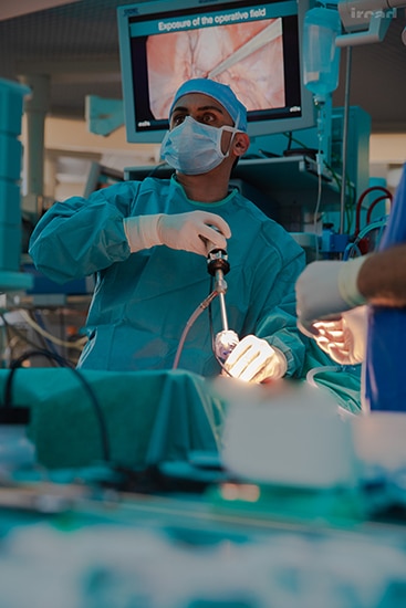
Dr. Aslam Noorbhai
Specialist Laparoscopic and Endoscopic Surgeon
- Phone:+27 31 582 5395
- Email:[email protected]

Dr Tendani Muthambi
Specialist General Surgeon

Dr Fusi Mosai
Specialist General Surgeon
- Phone: 066 474 1603
- Email:[email protected]

Dr Ignatius Botha
GASTROENTEROLOGY
- Phone:+27 (0)21 930 3014
- Email:[email protected]

Dr Morvendhran Moodley
Specialist Surgeon
- Phone:+27 (0)31 581 2376
- Email:[email protected]
Arthroscopic release of frozen shoulder
Arthroscopic Release of Frozen Shoulder Procedure: Medical Overview and Steps
Arthroscopic release of frozen shoulder, also known as arthroscopic capsular release or arthroscopic manipulation, is a minimally invasive surgical procedure performed to treat adhesive capsulitis, commonly known as frozen shoulder. This condition involves the gradual tightening and thickening of the joint capsule, leading to restricted shoulder movement and pain. Arthroscopic release aims to alleviate pain and improve shoulder mobility.
Procedure Steps:
Preoperative Assessment: Prior to surgery, the patient undergoes a comprehensive evaluation, including a review of medical history, physical examination, and imaging studies (such as X-rays and MRI) to assess the severity of the frozen shoulder and rule out other conditions.
Informed Consent: The patient is provided with detailed information about the procedure, potential risks, benefits, and alternative treatments. Informed consent is obtained, ensuring the patient’s understanding and agreement.
Anesthesia: The patient is typically placed under general anesthesia or regional anesthesia (nerve block) to ensure their comfort during the procedure.
Surgical Approach: Arthroscopic release is performed using a small, thin camera called an arthroscope and specialized surgical instruments. Several small incisions are made around the shoulder joint to insert the arthroscope and instruments.
Visualization and Capsular Release: The arthroscope provides a clear view of the interior of the shoulder joint on a monitor. The surgeon carefully identifies and releases the tight and thickened portions of the joint capsule that are limiting shoulder movement. This may involve cutting or releasing the adhesions using specialized instruments.
Closure and Recovery: After the procedure, the incisions are closed using sutures or adhesive strips. The patient is closely monitored as they wake up from anesthesia.
Postoperative Care: Pain management, physical therapy, and rehabilitation are essential components of recovery. The patient may need to wear a sling for a period to support the shoulder and facilitate healing.
Recovery and Rehabilitation: Physical therapy is crucial in the recovery process to regain shoulder mobility and strength. The therapist guides the patient through exercises and stretches to gradually restore full range of motion and function.
References:
American Academy of Orthopaedic Surgeons. (2021). Adhesive Capsulitis (Frozen Shoulder). Retrieved from https://orthoinfo.aaos.org/en/diseases–conditions/adhesive-capsulitis-frozen-shoulder
Mayo Clinic. (2021). Frozen Shoulder. Retrieved from https://www.mayoclinic.org/diseases-conditions/frozen-shoulder/diagnosis-treatment/drc-20372612
American Shoulder and Elbow Surgeons. (2021). Adhesive Capsulitis (Frozen Shoulder). Retrieved from https://www.orthopaedicsone.com/display/Main/Adhesive+Capsulitis
University of California, San Francisco (UCSF) Department of Orthopaedic Surgery. (2021). Arthroscopic Capsular Release for Adhesive Capsulitis (Frozen Shoulder). Retrieved from https://orthosurgery.ucsf.edu/patient-care/divisions/sports-medicine/conditions/shoulder/adhesive-capsulitis-frozen-shoulder.html
American Journal of Sports Medicine. (2019). Surgical Release for Frozen Shoulder: A Systematic Review and Meta-analysis of Clinical Trials. Retrieved from https://journals.sagepub.com/doi/abs/10.1177/0363546518781011
Please note that the specifics of arthroscopic release procedures for frozen shoulder may vary based on individual patient factors and the surgeon’s approach. Always consult with qualified orthopedic surgeons and healthcare professionals for personalized advice and information regarding arthroscopic release of frozen shoulder procedures.
Below is some Orthopaedic Surgeons that can assist with Arthroscopic Release of Frozen Shoulder Surgery

Dr Chris McCready
Orthopaedic Surgeon
- Phone:011 647 3571
- Email:[email protected]

Dr. P.H Laubscher
Specialist Orthopaedic Shoulder Surgeon
- Phone:(+27) 11 788-5110
- Email:[email protected]

Dr Shaun de Villiers
Orthopaedic Specialist
- Phone:043 422 0469
- Email:[email protected]

Dr Chris Narramore
Orthopedic ankle & shoulder specialist
- Phone:+27 21 762 5228
- Email:[email protected]

Dr Andries van Niekerk
Orthopaedic Surgeon
- Phone:012 664 2014
- Email:[email protected]

Dr Beyers Oosthuizen
Orthopaedic Surgeon
- Phone:+27 (0)12 652 9431
- Email:[email protected]

Dr Stephen Smith
Orthopaedic Surgeon
- Phone:(010) 786-4977
- Email:[email protected]

Dr Iwan Scott
Orthopaedic Surgeon
- Phone:015 291 5405
- Email:[email protected]
Arthroscopy
Arthroscopy Procedure: Medical Overview and Steps
Arthroscopy is a minimally invasive surgical procedure that allows a surgeon to visualize, diagnose, and treat various joint-related conditions using a specialized instrument called an arthroscope. This thin, flexible tube has a camera attached to it, which provides real-time images of the joint’s interior on a monitor. Arthroscopy is commonly used to examine and treat joint problems in the knee, shoulder, hip, wrist, ankle, and other joints.
Procedure Steps:
Preoperative Assessment: Before surgery, the patient undergoes a comprehensive evaluation, including a review of medical history, physical examination, and possibly imaging studies (X-rays, MRI, CT scans) to diagnose the joint issue and plan the arthroscopic procedure.
Informed Consent: The patient is provided with detailed information about the procedure, potential risks, benefits, and alternative treatments. Informed consent is obtained, ensuring the patient’s understanding and agreement.
Anesthesia: The patient is usually placed under regional anesthesia (nerve block) or general anesthesia to ensure their comfort during the procedure.
Surgical Approach: Small incisions (portals) are made near the joint to insert the arthroscope and other surgical instruments. Typically, two to four portals are used, depending on the joint being treated.
Visualization and Diagnosis: The arthroscope is inserted through one of the portals, and the joint’s interior is illuminated and magnified by the camera. The surgeon evaluates the structures within the joint, such as cartilage, ligaments, tendons, and bones, to diagnose any issues.
Treatment or Repair: Based on the diagnostic findings, the surgeon can perform various procedures using specialized instruments inserted through the other portals. Common arthroscopic procedures include removing damaged tissue (debridement), repairing torn ligaments or tendons, removing loose bodies, and smoothing damaged cartilage.
Closure and Recovery: After the procedure, the incisions are usually closed with sutures or adhesive strips. The patient is closely monitored as they wake up from anesthesia.
Postoperative Care and Rehabilitation: The recovery process varies depending on the joint and the procedure performed. Physical therapy is often recommended to promote healing, regain strength, and restore function. Patients are given instructions on wound care, pain management, and activity restrictions during recovery.
References:
American Academy of Orthopaedic Surgeons. (2021). Arthroscopy. Retrieved from https://orthoinfo.aaos.org/en/treatment/arthroscopy
Mayo Clinic. (2021). Arthroscopy. Retrieved from https://www.mayoclinic.org/tests-procedures/arthroscopy/about/pac-20392974
Cleveland Clinic. (2021). Arthroscopy. Retrieved from https://my.clevelandclinic.org/health/treatments/17933-arthroscopy
American Association of Orthopaedic Surgeons. (2021). Diagnostic and Operative Arthroscopy. Retrieved from https://www.orthopaedicsone.com/display/Main/Indications+for+Diagnostic+and+Operative+Arthroscopy
Journal of Bone and Joint Surgery. (2019). Practice Management Guidelines for Arthroscopy. Retrieved from https://journals.lww.com/jbjsjournal/fulltext/2019/09000/practice_management_guidelines_for_arthroscopy.23.aspx
Please note that the specifics of arthroscopy procedures may vary based on the joint being treated and the surgeon’s approach. Always consult with qualified orthopedic surgeons and healthcare professionals for personalized advice and information regarding arthroscopy procedures.
Below is some Orthopaedic Surgeons that can assit with Arthroscopy Surgery

Dr Beyers Oosthuizen
Orthopaedic Surgeon
- Phone:+27 (0)12 652 9431
- Email:[email protected]

Dr Andries van Niekerk
Orthopaedic Surgeon
- Phone:012 664 2014
- Email:[email protected]

Dr Shaun de Villiers
Orthopaedic Specialist
- Phone:043 422 0469
- Email:[email protected]

Dr. P.H Laubscher
Specialist Orthopaedic Shoulder Surgeon
- Phone:(+27) 11 788-5110
- Email:[email protected]

Dr Gabriel Pirjol
Orthopaedic Surgeon
- Phone:+27 31 202 5463
- Email:[email protected]

Dr. S.K Mafeelane
Specialist Orthopaedic Surgeon
- Phone:+27 15 880 1769
- Email:[email protected]

Dr. Peter Smith
Orthopaedic Surgeon
- Phone:+27 (0)21 552 2975
- Email:[email protected]

Dr Jan de Vos
Orthopaedic Surgeon
- Phone:012 807 0335
- Email:[email protected]
Assisted vaginal delivery (antenatal use)
Assisted vaginal delivery refers to a childbirth technique in which medical instruments are used to aid in the delivery of the baby, usually when the mother is unable to push the baby out on her own or when there are concerns for the baby’s well-being.
Here’s a general overview of the procedure:
Indications: Assisted vaginal delivery is indicated in cases where the mother is having difficulty pushing the baby out or when the baby is in distress. It can also be considered if the labor process has been prolonged, and maternal fatigue becomes a concern.
Instrumentation: Two common instruments used in assisted vaginal delivery are forceps and vacuum extractors. Forceps are tong-like instruments that are carefully applied to the baby’s head to help guide it through the birth canal. Vacuum extractors use suction to hold the baby’s head and assist in pulling it out.
Procedure: During the procedure, the obstetrician will assess the baby’s position and the mother’s progress in labor. If deemed appropriate, the chosen instrument (forceps or vacuum) will be carefully applied to the baby’s head. The obstetrician will guide and support the baby’s head as the mother continues to push during contractions. The instrument is used to provide gentle traction and guide the baby’s descent.
Monitoring: Throughout the procedure, the baby’s heart rate and the mother’s condition are closely monitored. If any complications or concerns arise, the medical team can quickly transition to other delivery methods, such as a cesarean section.
Recovery and Care: After the assisted vaginal delivery, the mother and baby will be closely monitored for any signs of complications. The mother’s perineal area (the tissue between the vagina and anus) may require stitches if it tears during delivery.
Here are five medical references you can explore for more in-depth information on assisted vaginal delivery:
American College of Obstetricians and Gynecologists (ACOG): Website: www.acog.org ACOG provides guidelines and resources related to obstetric practices, including assisted vaginal delivery techniques.
PubMed – National Library of Medicine: Website: www.ncbi.nlm.nih.gov/pubmed Search for scholarly articles and studies related to assisted vaginal delivery techniques and outcomes.
UpToDate: Website: www.uptodate.com UpToDate offers comprehensive articles on obstetric procedures, including assisted vaginal delivery and associated considerations.
Obstetrics & Gynecology Journal (The Green Journal): Website: journals.lww.com/greenjournal/pages/default.aspx This peer-reviewed journal publishes research and reviews related to obstetrics and gynecology, including articles about assisted vaginal delivery.
Mayo Clinic: Website: www.mayoclinic.org The Mayo Clinic provides patient-friendly information about childbirth procedures, including explanations of assisted vaginal delivery.
As always, it’s important to consult with healthcare professionals for personalized advice and recommendations regarding childbirth and delivery procedures.
Below is eight very reputable gynaecologists.

Dr Natalia Novikova
Gynaecologist and Endoscopic Surgeon
- Phone:076 242 0027
- Email: [email protected]

Dr Emma Bryant
General gynaecologist and gynaecologic oncologist
- Phone:+27 (0)12 346 2634
- Email:[email protected]

Dr Mutanda-Musoke
Specialist Obstetricians and Gynaecologists
- Phone:+27 (0) 12 003 4030
- Email:[email protected]

Dr Gitesh Rampersadh
Specialist Gynaecologist and Obstetrician
- Phone:+27 (0)31 265 1020
- Email:[email protected]

Dr Stephan Baloyi
Specialist Obstetrician and Gynaecologist
- Phone:(011) 432 4929
- Email:[email protected]

Dr Sandra Fonseca
specialist Obstetrician and Gynaecologist
- Phone:011 309 5900
- Email:[email protected]

Dr Paul Blaauwhof
Urogynaecologist/Endoscopic Surgeon
- Phone:010 035 0084
- Email:[email protected] <[email protected]
Axillary node clearance
Axillary node clearance, also known as axillary lymph node dissection, is a surgical procedure commonly performed as a part of breast cancer treatment. It involves the removal of lymph nodes from the axillary (armpit) region to determine the extent of cancer spread and to aid in staging and treatment planning.
Here’s a general overview of the procedure:
Indication: Axillary node clearance is often recommended when there is a suspicion or confirmation of cancer spread to the axillary lymph nodes. It helps in determining the stage of the cancer and guides further treatment decisions.
Surgical Procedure: The procedure is typically performed under general anesthesia. An incision is made in the axillary region, and the surgeon carefully dissects and removes the lymph nodes present. The number of nodes removed may vary depending on the extent of cancer spread and the surgeon’s judgment.
Pathological Examination: The removed lymph nodes are sent to a pathology laboratory for thorough examination. This examination helps in determining the extent of cancer involvement in the nodes and provides crucial information for treatment planning.
Recovery: After the surgery, patients may experience discomfort, swelling, and limited range of motion in the arm. Physical therapy and rehabilitation are often recommended to help regain normal arm function.
Potential Complications: As with any surgical procedure, axillary node clearance carries potential risks such as infection, bleeding, seroma formation (accumulation of fluid), nerve injury, and lymphedema (swelling due to compromised lymph drainage). However, the benefits of accurate staging and treatment planning generally outweigh the risks.
Here are five medical references you can explore for more in-depth information on axillary node clearance:
American Cancer Society: Website: www.cancer.org The American Cancer Society provides comprehensive information on various cancer-related topics, including axillary node clearance.
National Comprehensive Cancer Network (NCCN): Website: www.nccn.org The NCCN provides guidelines for cancer care, including recommendations for axillary node clearance in breast cancer treatment.
PubMed – National Library of Medicine: Website: www.ncbi.nlm.nih.gov/pubmed Search for scholarly articles and studies related to axillary node clearance. Use keywords like “axillary lymph node dissection” and “breast cancer.”
UpToDate: Website: www.uptodate.com UpToDate is a clinical decision support resource that offers detailed articles on medical procedures, including axillary node clearance.
Journal of Clinical Oncology: Website: ascopubs.org/journal/jco This peer-reviewed journal publishes research articles, reviews, and clinical studies related to oncology, including surgical procedures like axillary node clearance.
Remember that medical information can be complex, and it’s always best to consult with healthcare professionals and specialists for personalized advice and treatment recommendations.
Below is eight general Surgeons that can assist with Axillary node clearance surgery.

Dr Tendani Muthambi
Specialist General Surgeon

DR FATHIMA DOCRAT
General Surgeon
- Phone:+27 (0)11 951 0605/6
- Email:[email protected]

Dr Natasha Singh
Specialist General Surgeon
- Phone:27 (0)76 167 0982
- Email:Website from

Dr Gerhard Basson
General and Specialist surgeon
- Phone:012 652 9000
- Email:[email protected]

Dr Fusi Mosai
Specialist General Surgeon
- Phone: 066 474 1603
- Email:[email protected]

Dr Douglas Graham Ross
Specialist General Surgeon
- Phone:+2711 875 1992
- Email:[email protected]

DR AMISHA MARAJ
Specialist Surgeon
- Phone:067 944 8241
- Email:[email protected]

Dr Michael Heyns
GENERAL SURGEON
- Phone:+27(0)12 993 1160
- Email:[email protected]


